Hyperglycemia-Induced Oxidative Stress and Inflammatory Signaling in the Pathogenesis of Type 2 Diabetes Mellitus
Dr. Rachana Menon, Dr. Deepak Parchwani, Dr. Mehul Kaliya, Dr. Ragini Singh, Dr. Sagar Dholariya, Dr. Anita Motiani, Dr. Amit Sonagra
1 JR-2 (Acad) Department of Biochemistry, All India Institute of Medical Sciences, Rajkot
2Professor and Head, Department of Biochemistry, All India Institute of Medical Sciences, Rajkot
3Associate Professor, Department of General Medicine, All India Institute of Medical Sciences, Rajkot
4Additional Professor, Department of Biochemistry, All India Institute of Medical Sciences, Rajkot
5Associate Professor, Department of Biochemistry, All India Institute of Medical Sciences, Rajkot
APFCB News Volume 5, Issue 2
1. INTRODUCTION
Diabetes mellitus derives its name from the Greek diab?tes ("to pass through") and the Latin mellitus ("honey-sweet"), a nomenclature that reflects centuries of clinical observation predating modern biochemical characterization.[1,2] What was once identified purely by its cardinal symptom, the passage of abnormally sweet urine, is now recognized as a heterogeneous group of metabolic disorders unified by chronic hyperglycemia but distinguished by markedly different underlying mechanisms. Contemporary classification distinguishes the heterogenous etiologies of diabetes mellitus , ranging from the autoimmune destruction of pancreatic β-cells and absolute insulin deficiency in type 1 diabetes mellitus (T1DM), driven by autoimmune destruction of pancreatic β-cells and absolute insulin deficiency, the progressive insulin resistance and eventual insulin secretory failure in from type 2 diabetes mellitus (T2DM), characterized by progressive peripheral insulin resistance coupled with relative, and eventually absolute, insulin secretory failure. to transient gGestational diabetes and monogenic forms such as maturity-onset diabetes of the young account for a smaller, though clinically important, proportion of the overall disease burden. This etiological heterogeneity has significant implications for pathophysiological research, as mechanisms established in one form of diabetes cannot be assumed to generalize to another.
The global burden of diabetes has risen sharply over the past three decades. Worldwide prevalence increased from approximately 200 million affected individuals in 1990 to 830 million in 2022,[3] while contemporary estimates from the International Diabetes Federation indicate that 589 million adults currently live with diagnosed diabetes, with a further 252 million remaining undiagnosed. An additional 635 million adults have impaired glucose tolerance and 488 million have impaired fasting glucose, placing this population at substantially elevated risk of progression to overt disease. [4] This rise has not been uniform: the steepest increases in both prevalence and undiagnosed disease burden have occurred in low- and middle-income countries, where healthcare systems are often least equipped to manage the resulting long-term complications, underscoring diabetes as a disease of profound global health inequity rather than merely a consequence of affluence.
Type 2 diabetes mellitus accounts for approximately 90% of all diabetes cases and is defined by chronic hyperglycemia resulting from progressive resistance to the peripheral actions of insulin. Although historically considered a disease of middle and later life, epidemiological data now document a marked shift toward earlier age of onset.[5] Its pathogenesis reflects the interaction of non-modifiable risk factors, advancing age, family history, gestational diabetes, and specific ethnic backgrounds; with modifiable determinants including central obesity, physical inactivity, and dietary patterns.[5] Importantly, T2DM is no longer understood as a disorder confined to the classical triad of liver, muscle, and pancreas; contemporary pathophysiological models implicate a broader constellation of tissues, including adipose tissue, the gastrointestinal tract, kidney, brain, and vascular endothelium, each contributing distinct defects in glucose homeostasis, incretin signalling, and inflammatory tone. This expanding, multi-organ view of T2DM pathogenesis has reframed the disease as a systemic metabolic-inflammatory disorder rather than a purely glycemic one, a perspective central to the mechanistic focus of this review.
The clinical and economic consequences of T2DM are considerable. Rapid urbanization and economic transition have driven a global rise in disease burden,[6] with uncontrolled hyperglycemia predisposing to both macrovascular complications (coronary artery disease, cerebrovascular disease, peripheral arterial disease) and microvascular complications (retinopathy, neuropathy, nephropathy). Notably, subclinical vascular injury is frequently already present at the time of diagnosis, reflecting a preceding asymptomatic hyperglycemic interval of several years,[7] and cardiovascular disease remains the leading cause of mortality in this population. Encouragingly, early and intensive multifactorial intervention has been shown to substantially reduce complication risk, [8] with benefits that persist for years beyond the intervention period, a phenomenon termed the "legacy effect."[9] The persistence of this benefit, and conversely the persistence of harm from early poor control, points toward durable, tissue-level molecular changes that outlast the glycemic exposure that initiated them.
Despite considerable advances in glucose-lowering pharmacotherapy over the past two decades, the burden of diabetic complications remains disproportionately high relative to improvements in glycemic control alone, suggesting that hyperglycemia exerts its damage through mechanisms only partially addressed by glucose reduction itself. Oxidative stress and chronic low-grade inflammation have emerged as central, mechanistically interconnected pathways linking hyperglycemia to both insulin resistance and end-organ injury. This review examines the molecular mechanisms by which sustained hyperglycemia drives oxidative stress and inflammatory signaling, and how these interlinked pathways underlie the pathogenesis and progression of T2DM, with the aim of consolidating a fragmented mechanistic literature into a single, clinically oriented narrative framework.
2. CELLULAR AND MOLECULAR MECHANISMS OF INSULIN RESISTANCE IN TYPE 2 DIABETES
Insulin Synthesis and Secretion:
Insulin is a 51-residue anabolic peptide hormone synthesized as preproinsulin and processed through sequential proteolytic cleavage into its mature disulfide-linked A- and B-chain conformation within the secretory granules of pancreatic β-cells residing in the Islets of Langerhans. Nutrientstimulated insulin release is not a unitary event but a biphasic process, comprising an early, readily releasable pool of pre-docked granules followed by a sustained second phase dependent on granule mobilization and biosynthesis; this secretory dynamic is substantially potentiated by incretin hormones, principally glucagon-like peptide-1 (GLP-1) and gastric inhibitory polypeptide (GIP), which amplify glucose-stimulated insulin secretion through cAMP/PKA-dependent potentiation of β-cell exocytotic machinery.[10]
Insulin Receptor Structure and Downstream Signaling Pathways:
Insulin exerts its pleiotropic metabolic effects through the insulin receptor (IR), a disulfide-linked heterotetrameric (α?β?) member of the receptor tyrosine kinase superfamily, orchestrating glucose disposal predominantly in skeletal muscle and adipose tissue via GLUT4 transporter translocation, while concomitantly exerting suppressive control over hepatic gluconeogenic flux (Figure 1). Ligand engagement induces conformational activation and trans-autophosphorylation of the receptor's intracellular tyrosine kinase domains, generating phosphotyrosine docking motifs that recruit and phosphorylate insulin receptor substrate proteins, principally IRS-1 and IRS-2; alongside Shc adaptor proteins, thereby nucleating multiprotein signalling complexes.
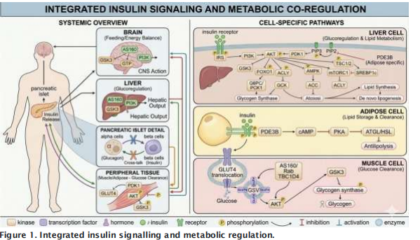
Figure 1. Integrated insulin signalling and metabolic regulation
Downstream signal propagation bifurcates into two functionally distinct cascades: the phosphoinositide 3-kinase (PI3K)/Akt axis (Figure 2), which subserves the metabolic and anabolic actions of insulin, and the Raf/Ras/MEK/mitogen-activated protein kinase (MAPK) pathway (Figure 3), which governs mitogenic, proliferative, and growth-regulatory programmes.[11]
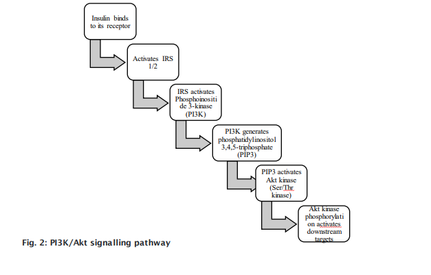
Fig. 3: Raf/Ras/MEK/MAPK signalling pathway
In T2DM, insulin-target tissues exhibit attenuated responsiveness to physiological hormone concentrations. This, a pathophysiological state designatesd insulin resistance., Itswhose molecular substrate is now understood to involves extensive lipotoxic and cytokine-mediated interference with proximal insulin signalling. Elevated circulating free fatty acids (FFAs), intracellular diacylglycerols (DAGs), and pro-inflammatory cytokines converge to disrupt canonical signal transduction at multiple nodes.
Tumor necrosis factor-alpha (TNF-α) drives this disruption by activatinges stress-responsive serine kinases, c-Jun N-terminal kinase (JNK) and IκB kinase-β (IKKβ), which catalyze aberrant inhibitory serine/threonine phosphorylation of IRS-1. This modification, sterically impairsing the tyrosinephosphorylation-dependent coupling of IRS-1 to the activated insulin receptor,its physiological tyrosine-phosphorylation-dependent coupling to the activated IRwhich subsequently blocks and abrogating Akt-mediated GLUT4 vesicle translocation.[12] Concurrently, the lipid phosphatase PTEN dephosphorylates the phosphoinositide second messenger PIP?, truncating the phosphoinositide gradient required for Akt membrane recruitment and activation. Meanwhile, , while adipocyte-derived interleukin-6 (IL-6) engages JAK-STAT signalling to induce suppressor of cytokine signalling (SOCS) proteins. These SOCS proteins, which target IRS-1/2 for ubiquitin-mediated proteasomal degradation.
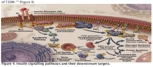
Together, these distinct mechanismshese mechanistically distinct yet convergent insults, serine/threonine hyperphosphorylation, phosphoinositide depletion, and substrate degradation, collectively attenuate flux through the PI3K/Akt axis at multiple hierarchical levels, establishing a durable, self-reinforcing state of cellular insulin resistance that constitutes the molecular substrate of T2DM.[13] (Figure 4)
3. ETIOPATHOGENESIS OF HYPERGLYCEMIA IN TYPE 2 DIABETES
When peripheral tissues develop insulin resistance, glucose disposal via GLUT4-mediated uptake becomes progressively impaired, shifting the burden of glycemic regulation onto hepatic glucose handling. Concurrent hepatic insulin resistance permits unrestrained transcriptional derepression of gluconeogenic enzymes, notably glucose-6-phosphatase and phosphoenolpyruvate carboxykinase (PEPCK), via impaired FoxO1 inhibition, thereby amplifying endogenous glucose output and establishing a self-perpetuating cycle of systemic hyperglycemia.[14] Pancreatic β-cells initially mount a compensatory response through hyperplastic and hypertrophic expansion of functional βcell mass, sustaining compensatory hyperinsulinemia sufficient to offset peripheral resistance. With prolonged exposure to chronic hyperglycemia, however, β-cells undergo progressive desensitization to glucose, a maladaptive state termed glucotoxicity, characterized by transcriptional downregulation of the glycemic sensor glucokinase and consequent attenuation of glucosestimulated insulin secretion (GSIS). Sustained hyperglycemia additionally suppresses mitochondrial fatty acid β-oxidation through malonyl-CoA-mediated allosteric inhibition of carnitine palmitoyltransferase-1 (CPT-1), redirecting excess lipid substrate into non-oxidative esterification pathways and promoting intracellular diacylglycerol (DAG) accumulation, a lipotoxic intermediate implicated in further signalling dysfunction.
The resultant glucolipotoxic microenvironment precipitates endoplasmic reticulum (ER) stress and activation of the unfolded protein response, culminating in transcriptional downregulation of the master insulin gene regulators pancreatic duodenal homeobox-1 (PDX-1) and V-maf musculoaponeurotic fibrosarcoma oncogene homolog A (MAFA), both indispensable for maintaining β-cell identity and insulin biosynthetic capacity. Compounded by locally elevated pro-inflammatory cytokines, tumor necrosis factor-alpha (TNF-α) and interleukin-1β (IL-1β), arising from islet-resident macrophage activation, these convergent glucolipotoxic and inflammatory insults drive progressive, mechanistically interdependent β-cell dedifferentiation and apoptotic attrition. This cascade culminates in the decompensated phase of T2DM, marked by an irreversible decline in functional βcell mass and secretory reserve, wherein compensatory hyperinsulinemia can no longer be sustained and overt, clinically manifest hyperglycemia ensues.[15]
4. HYPERGLYCEMIA-INDUCED OXIDATIVE STRESS AND METABOLIC DYSFUNCTION
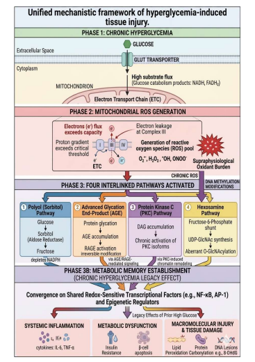
Oxidative stress is classically conceptualized as a pathological imbalance between the generation of reactive oxidant species and the buffering capacity of endogenous antioxidant defences, culminating in disrupted redox signalling and cumulative macromolecular injury. [16] Under physiological conditions, glucose undergoes catabolism via the Embden-Meyerhof-Parnas glycolytic pathway, generating the reducing equivalents NADH and FADH?, which donate electrons to complexes I and II of the mitochondrial electron transport chain to drive chemiosmotic ATP synthesis through oxidative phosphorylation.[17] During sustained hyperglycemia, however, excess substrate flux into the electron transport chain elevates the mitochondrial proton gradient beyond a critical threshold, inducing electron leakage at complex III and consequent overproduction of reactive oxygen species (ROS), including the hydroxyl radical (OH•), superoxide anion (O??•), hydrogen peroxide (H?O?), and peroxynitrite (ONOO?), the latter arising from the diffusion-limited reaction between superoxide and nitric oxide. Although enzymatic (superoxide dismutase, glutathione peroxidase, catalase) and nonenzymatic (vitamins A, C, and E, reduced glutathione) antioxidant systems ordinarily neutralize physiological ROS flux, the supraphysiological oxidant burden characteristic of T2DM exceeds this buffering capacity, precipitating a sustained state of oxidative stress. [16] This unchecked ROS accumulation inflicts direct macromolecular injury, peroxidation of membrane lipids, oxidative carbonylation of structural and enzymatic proteins, and formation of mutagenic DNA lesions such as 8-hydroxy-2′-deoxyguanosine, while simultaneously functioning as a proximal trigger for four interlinked, mechanistically distinct pro-oxidative pathways that amplify and perpetuate cellular injury: the polyol pathway, advanced glycation end-product formation and receptor engagement, protein kinase C isoform activation, and hexosamine biosynthetic pathway flux. Rather than operating in isolation, these pathways converge on shared downstream mediators, most notably further ROS generation and redox-sensitive transcriptional activation, establishing oxidative stress as both a consequence and a self-amplifying driver of hyperglycemia-induced tissue injury in T2DM.[18]
4.1 The Polyol (Sorbitol) Pathway
The polyol pathway constitutes an accessory, NADPH-dependent route of glucose catabolism operative chiefly in tissues exhibiting insulin-independent glucose uptake, the lens, retina, peripheral nerves, kidney, and seminal vesicles, rendering these sites selectively vulnerable to hyperglycemic substrate flooding independent of insulin-mediated glucose transport regulation. The rate-limiting enzyme aldose reductase, a member of the NADPH-dependent aldo-keto reductase superfamily, reduces glucose to sorbitol, which is subsequently oxidized to fructose by sorbitol dehydrogenase in an NAD?-dependent reaction.[17] Under euglycemic conditions, aldose reductase exhibits low affinity for glucose relative to its physiological substrates and thus contributes negligibly to overall glucose disposal; however, during sustained hyperglycemia, saturation of the high-capacity glycolytic and hexokinase-mediated pathways diverts substantial glucose flux into this otherwise quiescent route. This shunting imposes a critical metabolic cost: aldose reductase-mediated glucose reduction consumes NADPH stoichiometrically, thereby depleting the cofactor pool required for glutathione reductase-mediated regeneration of reduced glutathione, the principal intracellular thiol antioxidant, and compounding the prevailing oxidative burden. Because sorbitol is a polar, poorly membranepermeant polyol and is further metabolized inefficiently in tissues with limited sorbitol dehydrogenase expression, its intracellular accumulation generates a hypertonic gradient that drives osmotic water influx, cellular swelling, and structural injury. This osmotic-oxidative dual insult underlies several hallmark microvascular complications of diabetes, manifesting clinically as diabetic cataract formation, macular edema, retinopathy, peripheral neuropathy, and nephropathy, and positions the polyol pathway as a mechanistic nexus linking hyperglycemia, redox imbalance, and tissue-specific microvascular injury.[18]
4.2 Advanced Glycation End-Products (AGEs) and Receptor Activation (RAGE)
Advanced glycation end-products (AGEs) arise via the Maillard reaction, an irreversible, nonenzymatic cascade of cross-linking condensation reactions between reducing sugars and the nucleophilic amine groups of proteins, lipids, or nucleic acids, proceeding through unstable Schiff base and Amadori intermediates toward stable, heterogeneous AGE adducts, a process that occurs at a low, physiologically tolerable basal rate under euglycemic conditions.[17] Sustained hyperglycemia substantially accelerates the kinetics of AGE formation beyond the capacity of receptor-mediated and proteolytic clearance mechanisms, resulting in progressive tissue accumulation within the vasculature, renal parenchyma, peripheral nerves, ocular structures, and long-lived collagen matrices, the latter rendering AGE burden particularly cumulative given collagen's slow turnover. AGEs exert their pathogenic effects principally through engagement of RAGE (receptor for advanced glycation end-products), a multiligand pattern-recognition receptor of the immunoglobulin superfamily broadly expressed across cardiac, pulmonary, skeletal muscle, and vascular tissue, as well as neuronal, glial, endothelial, and innate immune cell populations. AGE-RAGE ligation triggers receptor dimerization and downstream activation of redox-sensitive transcription factors, including nuclear factor kappa B (NF-κB), activator protein-1 (AP-1), and forkhead box protein O4 (FoxO4), thereby propagating a self-sustaining signalling loop of inflammatory gene transcription, vascular endothelial injury, cellular dysfunction, and amplified ROS generation. Cross-linking of AGEs within structural vascular proteins, notably collagen and elastin, imparts pathological arterial stiffening and impaired vasoreactivity, promoting atherosclerotic plaque development, coronary artery disease, and cerebrovascular events, while tissue-resident AGE accumulation independently and synergistically contributes to the microvascular pathology underlying diabetic retinopathy, nephropathy, and peripheral neuropathy.[19]
4.3 Activation of Protein Kinase C (PKC) Isoforms
Protein kinase C (PKC) constitutes a structurally diverse family of serine/threonine kinases, comprising conventional (calcium- and DAG-dependent), novel (calcium-independent, DAGdependent), and atypical isoforms, each regulating distinct downstream substrate networks through phosphorylation-dependent conformational activation. Under physiological glycolytic flux, glyceraldehyde-3-phosphate, a triose phosphate intermediate generated by aldolase-mediated cleavage of fructose-1,6-bisphosphate — exists in reversible enzymatic equilibrium with its structural isomer dihydroxyacetone phosphate via triose phosphate isomerase, the latter serving as the obligate precursor for glycerol-3-phosphate synthesis and, by extension, the glycerol backbone of triglycerides and membrane phospholipids. During sustained hyperglycemia, however, glycolytic substrate loading shifts this equilibrium toward dihydroxyacetone phosphate accumulation, driving its enzymatic reduction to glycerol-3-phosphate and subsequent de novo synthesis of diacylglycerol (DAG), a lipid second messenger and the principal endogenous allosteric activator of conventional and novel PKC isoforms. Hyperglycemia-induced DAG accumulation thus produces sustained, pathological PKC activation, in contrast to the transient, tightly regulated activation characteristic of physiological signal transduction. Once activated, PKC isoforms, particularly PKC-β and PKC-δ, phosphorylate a broad array of downstream effectors implicated in vascular pathology, including NADPH oxidase subunits, endothelial nitric oxide synthase, and components of the mitochondrial electron transport chain, thereby further augmenting ROS generation. This establishes a selfpropagating feed-forward loop in which hyperglycemia-driven DAG-PKC signalling both originates from and perpetuates oxidative injury, contributing mechanistically to the vascular permeability defects, basement membrane thickening, and hemodynamic abnormalities characteristic of diabetic microvascular complications.[20]
4.4 Hexosamine Pathway Flux
The hexosamine biosynthetic pathway (HBP) constitutes a nutrient-sensing metabolic offshoot that diverts a minor fraction of glycolytic flux away from glycolysis proper, supporting the biosynthesis of uridine diphosphate N-acetylglucosamine (UDP-GlcNAc), the obligate substrate for N-linked and Olinked (O-GlcNAcylation) glycosylation reactions that dynamically regulate protein stability, subcellular localization, transcriptional activity, and intracellular signal transduction. Under physiological conditions, the rate-limiting enzyme glutamine:fructose-6-phosphate amidotransferase (GFAT) catalyzes the committed step of the pathway, converting fructose-6-phosphate and glutamine to glucosamine-6-phosphate, which is subsequently processed through a series of acetylation, isomerization, and uridylation reactions to yield UDP-GlcNAc, thereby coupling cellular nutrient status to protein post-translational modification. During sustained hyperglycemia, however, glycolytic substrate excess drives transcriptional and enzymatic upregulation of GFAT, disproportionately elevating flux through the HBP and generating supraphysiological UDP-GlcNAc pools. This excess substrate availability promotes aberrant O-GlcNAcylation of key transcriptional regulators, including Sp1, resulting in increased expression of transforming growth factor -α and -β (TGF-α, TGF-β) and plasminogen activator inhibitor-1 (PAI-1). Upregulated TGF-β signalling suppresses mesangial cell mitogenesis while concurrently promoting extracellular matrix accumulation, mesangial expansion, and glomerular basement membrane thickening, structural alterations that are pathological hallmarks of early diabetic nephropathy. More broadly, hexosamine pathway -driven OGlcNAcylation interferes with insulin signalling by competitively modifying serine/threonine residues otherwise targeted for phosphorylation, thereby establishing a further mechanistic link between glucose-flux-dependent post-translational modification, extracellular matrix dysregulation, and the vascular, renal, and neural injury characteristic of chronic hyperglycemic exposure. [20]
4.5 Metabolic Memory: Persistent Injury Beyond Glycemic Correction
Beyond these four canonical pathways lies the phenomenon of "metabolic memory," a paradigm - shifting concept establishing that a preceding period of chronic hyperglycemic exposure confers durable, self-sustaining tissue injury that continues to drive microvascular and macrovascular complication progression despite subsequent attainment of near-normoglycemic control, a finding first substantiated by extended post-trial follow-up of intensively versus conventionally treated cohorts, wherein early glycemic divergence produced complication risk trajectories that persisted for years after glycemic parity was achieved between groups. Mechanistically, this persistence is attributed to the convergence of several self-perpetuating processes: sustained mitochondrial ROS overproduction that outlasts the initiating hyperglycemic stimulus, ongoing accumulation of irreversibly cross-linked, slowly-turned-over AGEs within long-lived structural proteins, and, most notably, epigenetic reprogramming of the cellular transcriptional landscape. This epigenetic dimension encompasses aberrant DNA methylation at promoter regions of key metabolic and inflammatory genes, altered histone methylation marks (notably H3K4 and H3K9 methylation at the NF-κB p65 promoter), and dysregulated histone acetylation, collectively producing a chromatin configuration that "locks" pro-inflammatory and pro-oxidative gene expression programmes, including NF-κB-responsive genes, in a constitutively activated, transcriptionally permissive state that is resistant to subsequent normalization of glycemia. Because these epigenetic marks are heritable across cell divisions and only slowly reversible, metabolic memory effectively converts a transient physiological exposure into a durable pathological trait embedded within the cellular transcriptional apparatus. This mechanistic understanding constitutes compelling biological rationale for early, intensive glycemic intervention at the point of diagnosis, rather than delayed, stepwise, or incremental treatment escalation, positioning the earliest phase of disease management as a critical, time-limited therapeutic window.[21] .
5. MOLECULAR CROSSTALK: FROM OXIDATIVE STRESS TO CHRONIC INFLAMMATION
Oxidative stress and inflammation are increasingly recognized as mechanistically interdependent, bidirectionally coupled processes rather than discrete, sequentially ordered pathological events, reflecting a shared evolutionary origin in innate cellular stress-response signalling. Reactive oxygen species promote inflammatory activation through both direct and indirect mechanisms: directly, via oxidative modification of membrane phospholipids and release of lipid-derived inflammatory mediators such as oxidized low-density lipoprotein and eicosanoids; and indirectly, through redox-sensitive activation of a broad transcriptional network encompassing peroxisome proliferator -activated receptorgamma (PPAR-γ), nuclear factor kappa B (NF-κB), activator protein-1, p53, nuclear factor erythroid 2- related factor 2 (Nrf2), and hypoxia-inducible factor-1α (HIF-1α), several of which exhibit reciprocal cross-regulation that further amplifies the inflammatory transcriptional output. Among these, NF-κB occupies a position of particular mechanistic centrality, constituting a family of structurally related, inducible transcription factors, including the canonical p65/p50 heterodimer, that govern innate and adaptive immunity, inflammatory gene expression, cell survival, and proliferative signalling. Under quiescent basal conditions, NF-κB dimers are held in transcriptionally inert cytoplasmic sequestration through stoichiometric association with their inhibitory protein, IκB, which masks the nuclear localization sequence and thereby prevents nuclear entry. ROS-mediated activation of the IκB kinase (IKK) complex catalyzes phosphorylation-dependent ubiquitination and proteasome degradation of IκB, liberating NF-κB for nuclear translocation and subsequent transactivation of a broad inflammatory gene programme encoding interleukin-1 (IL-1), interleukin-6 (IL-6), and tumor necrosis factor-alpha (TNF-α). The resulting cytokine milieu drives endothelial dysfunction through impaired nitric oxide bioavailability, promotes vascular smooth muscle cell proliferation and migration, and accelerates atherogenic plaque formation, thereby mechanistically linking redox dysregulation to the macro vascular complications of chronic hyperglycemia.
Critically, this inflammatory activation does not remain confined to vascular tissue but directly reinforces the insulin-resistant phenotype at the level of proximal insulin signalling, establishing a pathogenic convergence between the inflammatory and metabolic disease axes. Activated IKK-β, in addition to its canonical role in NF-κB liberation, exerts a moonlighting kinase function by directly phosphorylating insulin receptor substrate-1 and -2 (IRS-1/2) on inhibitory serine residues, a modification that stands in direct mechanistic opposition to the physiological tyrosine phosphorylation induced by ligand-activated insulin receptor auto phosphorylation. Because serine-phosphorylated IRS-1/2 exhibits diminished capacity to couple productively with the activated insulin receptor and to nucleate downstream phosphoinositide 3-kinase (PI3K)/Akt signalling complexes, this post-translational modification functions as a molecular switch that actively uncouples insulin receptor activation from its physiological metabolic output. Consequently, this reciprocal cross-talk establishes a self-amplifying, feed-forward pathological cycle in which oxidative stress drives NF-κB/IKK-β-dependent inflammatory activation, inflammatory signalling in turn deepens insulin resistance through serine-phosphorylation-mediated IRS-1/2 inactivation, and the resulting metabolic dysregulation generates further substrate-driven ROS overproduction — collectively constituting a mechanistically closed, self-perpetuating circuit that sustains and progressively intensifies the pathophysiological trajectory of type 2 diabetes mellitus. [22]
6. CONCLUSION AND FUTURE PERSPECTIVES
The convergence of insulin signalling defects, hyperglycemia-driven oxidative stress, and NF-κBmediated inflammatory activation forms a self-reinforcing molecular circuit that underlies both the initiation and progression of T2DM and its vascular complications. The phenomenon of metabolic memory reinforces the clinical principle that early, intensive glycemic control is essential to limit the durable, epigenetically encoded tissue injury that persists even after glycemic targets are subsequently achieved.
Translating this mechanistic understanding into clinical benefit will require several parallel advances: the development of accessible, non-invasive biomarkers (e.g. urinary 8-OHdG and MDA; salivary AGEs, CRP, and IL-6; urinary or salivary exosomal miRNAs) that more accurately reflect real-time insulin sensitivity, β-cell function, and oxidative burden; targeted screening strategies for earlier identification of at-risk individuals; and public health interventions promoting dietary modification and regular aerobic activity as first-line, modifiable interventions. Furthermore, since oxidative injury begins early on, future research must prioritise targeted screening as well as precision medicine framework approaches (individualised therapies, molecular phenotyping) integrated with machine learning algorithms, to intercept well before the occurrence of irreversible tissue damage. Emerging antioxidant- and antiinflammatory-targeted therapeutics, alongside conventional glucose-lowering agents – most notably GLP-1 receptor agonists and SGLT2 inhibitors -, warrant continued investigation as adjuncts capable of interrupting the oxidative-inflammatory cycle described in this review. Apart from glycemic control, their well-established cardio-protective and reno-protective effects strongly support their ability to directly mitigate mitochondrial injury, oxidative stress, chronic inflammation and endothelial dysfunction in T2DM. A deeper understanding of the interplay between insulin resistance, insulin secretory capacity, and redox and inflammatory signalling remains central to the development of more effective, mechanistically informed therapeutic strategies for T2DM.
1. Sun H, Saeedi P, Karuranga S, et al. IDF Diabetes Atlas: global, regional and country -level diabetes prevalence estimates for 2021 and projections for 2045. Diabetes Res Clin Pract. 2022;183:109119.
2. Poretsky L. Milestones in the history of diabetes mellitus. World J Diabetes. 2016;7(1):1–7.
3. NCD Risk Factor Collaboration (NCD-RisC). Worldwide trends in diabetes prevalence and treatment from 1990 to 2022: a pooled analysis of 1108 population-representative studies with 141 million participants. Lancet. 2024;404(10468):2125–2143.
4. International Diabetes Federation. IDF global clinical practice recommendations for managing type 2 diabetes—2025 [Internet]. Brussels: IDF; 2025 [cited 2026 Jul 9]. Available from: https://idf.org/t2d-cpr-2025 5. Lascar N, Brown J, Pattison H, et al. Type 2 diabetes in young adults: challenges and future possibilities. Lancet Diabetes Endocrinol. 2018;6(1):69–80.
6. Bommer C, Sagalova V, Heesemann E, et al. Global economic burden of diabetes in adults: projections from 2015 to 2030. Diabetes Care. 2018;41(5):963–970.
7. Parchwani DN, Murthy SMS, Upadhyah AA, Patel DD. Genetic factors in the etiology of type 2 diabetes: linkage analyses, candidate gene association, and genome-wide association – still a long way to go! Natl J Physiol Pharm Pharmacol 2013; 3:57-68.
8. Gaede P, Oellgaard J, Carstensen B, et al. Years of life gained by multifactorial intervention in patients with type 2 diabetes mellitus and microalbuminuria: 21 years of follow-up on the Steno2 randomised trial. Lancet Diabetes Endocrinol. 2016;4(12):1008–1016.
9. halmers J, Cooper ME. UKPDS and the legacy effect. N Engl J Med. 2008;359(15):1618–1620.
10. Yamanouchi D. The roles of incretin hormones GIP and GLP-1 in metabolic and cardiovascular health: a comprehensive review. Int J Mol Sci. 2026;27(1):27.
11. Martínez Báez A, Ayala G, Pedroza-Saavedra A, González-Sánchez HM, Chihu Amparan L. Phosphorylation codes in IRS-1 and IRS-2 are associated with the activation/inhibition of insulin canonical signaling pathways. Curr Issues Mol Biol. 2024;46(1):41.
12. Woo JR, Bae SH, Wales TE, et al. The serine phosphorylations in the IRS-1 PIR domain abrogate IRS-1 and IR interaction. Proc Natl Acad Sci U S A. 2024;121(17):e2401716121.
13. Ueki K, Kondo T, Tseng YH, Kahn CR. Central role of suppressors of cytokine signaling proteins in hepatic steatosis, insulin resistance, and the metabolic syndrome in the mouse. Proc Natl Acad Sci U S A. 2004;101(28):10422–10427.
14. Onyango AN. Excessive gluconeogenesis causes the hepatic insulin resistance paradox and its sequelae. Heliyon. 2022;8(12):e12294. 15.
15. Poitout V, Robertson RP. Glucolipotoxicity: fuel excess and β-cell dysfunction. Endocr Rev. 2008;29(3):351–366. 16.
16. Sies H. Oxidative stress: concept and some practical aspects. Antioxidants (Basel). 2020;9(9):852. 17.
17. Wani ZA, Sharma AK, Muzamil S, et al. Molecular pathways of oxidative stress in diabetes: redox imbalance and insulin pathway dysregulation. Mol Biol Rep. 2026;53:222. 18.
18. Brownlee M. Biochemistry and molecular cell biology of diabetic complications. Nature. 2001;414(6865):813–820. 19.
19. Yamagishi S, Nakamura K, Matsui T, Noda Y, Imaizumi T. Receptor for advanced glycation end products (RAGE): a novel therapeutic target for diabetic vascular complication. Curr Pharm Des. 2008;14(5):487–495. 20.
20. Parchwani DN, Vachhani U, Dholariya S, Agravatt A, Singh R, Sonagra A. Role of Inflammatory and Oxidative Stress Biomarkers with Albuminuria: A Cross Sectional Analysis in Type 2 Diabetes Mellitus Patients. EJIFCC. 2025 Oct 1;36(3):284-297. 21.
21. Ceriello A, Lucisano G, Prattichizzo F, et al. The legacy effect of hyperglycemia and early use of SGLT-2 inhibitors: a cohort study with newly-diagnosed people with type 2 diabetes. Lancet Reg Health Eur. 2023;31:100666. 22.
22. Liu T, Zhang L, Joo D, Sun SC. NF-κB signaling in inflammation. Signal Transduct Target Ther. 2017;2:17023.