Protein Electrophoresis Patterns in Orbital Apex Syndrome: A Case Series of Rare Diagnosis
Dr. Swati Singh, Dr Manushri Sharma, Dr Bhawna Mahajan, Dr Jitender Sharma, Dr Renu Sehrawat, Dr Cheteny Cheteny, Dr Mohit Singh
Department of Biochemistry, Govind Ballabh Pant Institute of Postgraduate Medical Education and Research, New Delhi, India
APFCB News Volume 5, Issue 2
Introduction Orbital Apex Syndrome (OAS) is a rare neuro-ophthalmic disorder caused by lesions involving the orbital apex, resulting in dysfunction of the optic nerve and cranial nerves III, IV, VI, and the ophthalmic division of the trigeminal nerve [1] . Clinical features include ophthalmoplegia, proptosis, ptosis, hypoesthesia of the forehead, and vision loss. OAS has a multifactorial etiology and may arise secondary to neoplastic, infectious, inflammatory, traumatic, or vascular disorders, including plasma cell dyscrasias, systemic lupus erythematosus, Tolosa–Hunt syndrome, bacterial or fungal sinusitis, herpes zoster, iatrogenic injury, craniomaxillofacial fractures, and cavernous sinus thrombosis [1,2] .
Given the risk of irreversible visual loss, permanent cranial neuropathies, intracranial spread, and potentially life-threatening complications, OAS requires prompt diagnosis and treatment. However, its heterogeneous etiologies, nonspecific clinical manifestations, and overlap with other orbital and neuro-ophthalmic disorders often make early diagnosis challenging. Current evidence remains largely confined to isolated case reports and small case series, with limited data to inform standardized diagnostic strategies or the incorporation of adjunctive investigations, such as serum protein electrophoresis (SPE), into routine clinical practice [3-5]. While contrast-enhanced CT and MRI accurately localize orbital apex lesions and define their extent, they often do not establish the underlying systemic diagnosis, particularly in infiltrative or hematological disorders.Thus, Although imaging and histopathological examination remain the cornerstone of OAS diagnosis by localizing the orbital lesion, they often fail to identify the underlying systemic etiology [6]. In contrast, the diagnostic utility of laboratory investigations, particularly SPE, remains poorly characterized and is seldom discussed in the literature despite its potential to detect plasma cell dyscrasias and other systemic disorders that may underlie OAS. It is important because plasma cell dyscrasias and other monoclonal gammopathies may initially present with orbital involvement, inexpensive laboratory investigations such as SPE may provide an important diagnostic clue before systemic manifestations become clinically apparent [7]. We present a case series of three patients with OAS illustrating distinct underlying etiologies, highlighting the use of serum protein electrophoresis as an adjunctive investigation.
Case Reports
Case 1
A 45-year-old female patient presented with a 4-day history of right eye pain associated with blurred vision in both eyes. On examination, visual acuity was limited to finger counting at 1 m in both eyes. Extraocular movements were restricted in lateral and upward gaze in the right eye and in upward gaze in the left eye. Sensation was reduced in the left V1 distribution, while motor examination revealed no focal neurological deficits.
The laboratory, serological, and neuroimaging findings for all cases are detailed in Table 1. Complete blood count was within normal limits. Serum C-reactive protein (CRP), rheumatoid factor (RF), Hepatitis B surface antigen (HBsAg) and Hepatitis C antibodies (HCV) were negative. SPEP revealed polyclonal hypergammaglobulinemia (Figure 1B). Serum IgG4 was normal (0.94 g/L). Cerebrospinal fluid (CSF) analysis was unremarkable. Cartridge-Based Nucleic Acid Amplification Test (CB-NAAT) of the CSF was negative. Contrast-enhanced MRI of the orbits failed to demonstrate a localizing lesion. CECT brain and orbit revealed no significant abnormality. Ultrasonography (USG) of the whole abdomen and CECT of the chest and abdomen were unremarkable. Diagnosis of acute orbital apex syndrome was made clinically. The patient received intravenous immunoglobulin (IVIG), with subsequent clinical improvement.
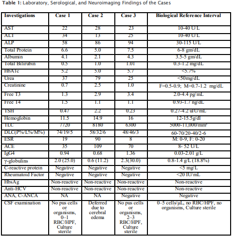
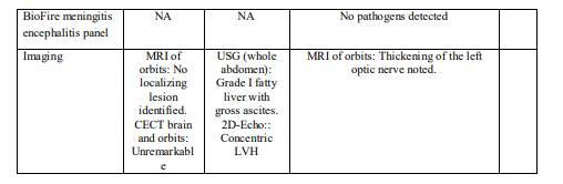
AST:Aspartate Aminotransferase; ALT: Alanine Aminotransferase; ALP: Alkaline Phosphatase; ANA:Antinuclear antibody; TSH: Thyroid Stimulating Hormone; ESR: Erythrocyte Sedimentation Rate; ACE: angiotensin-converting enzyme; HbsAg:Hepatitis B surface antigen; anti-HCV: Hepatitis C virus antibody; RBC: Red blood cells; HPF: High power field; NA: Not available; TLC:total leukocyte count; 2D Echo: 2D echocardiography: LVH:left ventricular hypertrophy; USG: Ultrasonography
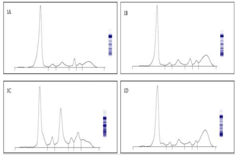
Figure 1A Normal serum electrophoretic pattern. 1B Case 1: SPEP revealed polyclonal hypergammaglobulinemia suggestive of chronic inflammatory or autoimmune etiology. 1C Case 2: SPEP demonstrated increased α2 and β2 fractions with distortion in β2 and γ. 1DCase 3: SPEP showed hypergammaglobulinemia with faint β1-region distortion, likely reflecting β-region immunoglobulin migration.
Case 2
A 54-year-old man presented with a 3.5-month history of right frontotemporal throbbing headache, lasting 2–3 hours and worsening in the evening. Eight days later, he developed binocular diplopia, right eye esotropia, proptosis, restricted ocular movements, and bilateral periorbital swelling. The headache subsequently recurred once or twice weekly. Examination revealed visual acuity of 6/24 (right) and 6/12 (left), anisocoria (right
Case 3
A 45-year-old male, tobacco chewer with no known comorbidities, presented with 1 month history of holocranial headache followed by horizontal diplopia and mild diminution of vision in the left eye. The diplopia shows maximum separation of images on left gaze, suggesting left lateral rectus weakness. Neurological examination revealed isolated left abducens palsy, with visual acuity 6/6 in the right eye and 6/9 in the left eye with mild impairment of color vision, while pupils, visual fields, and the rest of cranial nerves were normal. Motor, sensory, cerebellar, and systemic examinations were unremarkable, and there were no meningeal signs or long-tract deficits.
CBC revealed a TLC of 6,300/mm³. Serum ACE was 70 U/L, and IgG4 was 1.36 g/L. Serological testing was negative for HBsAg and anti-HCV, while anti-HBc was positive. Antinuclear antibody (ANA) testing by indirect immunofluorescence was negative. CSF microscopy and culture were unremarkable, with negative AFB smear. CSF cytology was negative for malignant cells (Table 1). Bacterial and fungal cultures, as well as the BioFire® FilmArray® Meningitis/Encephalitis (ME) Panel, were negative. SPEP showed hypergammaglobulinemia with faint β1-region distortion. MRI brain showed thickening of the left optic nerve.
Discussion
Serum protein electrophoresis is a simple, reliable, and accurate technique used to separate serum proteins [8]. The present study findings identified distinctive pathological electrophoretic patterns observed in three rare cases of OAS: one patient showed a pattern indicative of acute inflammatory response, one exhibited polyclonal hypergammaglobulinemia and one with chronic granulomatous process.
The first case involved a 45-year-old woman presenting with clinical features consistent with OAS. The diagnosis of OAS was made only after correlating the clinical picture with SPEP, CSF analysis, and a negative autoimmune workup, while initial brain and orbital imaging was inconclusive and failed to demonstrate definitive localizing findings. Further, In the context of etiology, in our patient, SPE demonstrated polyclonal hypergammaglobulinemia (2 g/L), a pattern suggestive of an underlying inflammatory or autoimmune condition due to increased immunoglobulin production, predominantly IgG type [7]. Previous reports have identified IgG4-related disease as a recognized autoimmune cause of OAS, albeit, normal serum IgG4 levels made this diagnosis less likely in our patient [9]. Nevertheless, polyclonal hypergammaglobulinemia and the favorable response to intravenous immunoglobulin support an underlying immune-mediated etiology unrelated to IgG4-related disease.
In our second case, SPE revealed a predominantly acute-phase reactant profile, with elevated α1- globulin (0.3 g/L) and α2-globulin (1.4 g/L) fractions, a normal γ-globulin fraction (0.6 g/L), and hypoalbuminemia (1.8 g/L). During acute inflammatory states, increased hepatic synthesis of acutephase reactants elevates the α1- and α2-globulin fractions, while cytokine-mediated suppression of albumin synthesis results in hypoalbuminemia [7]. Albeit, for this patient, elevated ACE level also raised the possibility of an underlying granulomatous or systemic/hepatic condition. Additionally, a normal CBC and absence of monoclonal protein on SPE excluded plasma cell dyscrasia–associated etiologies. Together, these findings explain the characteristic inflammatory SPE profile observed in our patient, supporting an inflammatory origin.
For the third case, SPEP demonstrated hypergammaglobulinemia with faint β1-region distortion. Hypergammaglobulinemia reflects increased immunoglobulin production associated with chronic inflammation, autoimmune, granulomatous, or chronic infectious conditions. The faint β1-region distortion is consistent with polyclonal immunoglobulin migration [7]. In the setting of negative infectious studies, negative ANA, normal serum IgG4 levels, and MRI findings of optic nerve thickening, these findings were compatible with an inflammatory granulomatous process, supporting the provisional diagnosis of neurosarcoidosis [10] . The cases in this series reflect the diagnostic heterogeneity of OAS. Case 1 lacked biopsy confirmation, Case 2 relied on a referral diagnosis, and Case 3 remained provisional neurosarcoidosis. This variability mirrors the diagnostic challenges encountered in clinical practice, where obtaining definitive tissue diagnosis is often challenging because of the anatomical location and the urgency of initiating treatment. Further, the collective findings of this case series illustrate that SPE provided valuable information by identifying distinct electrophoretic patterns of underlying inflammatory, vascular, and immune-mediated processes when conventional imaging alone was insufficient to establish the systemic etiology. Accordingly, the SPE findings should be interpreted as supportive rather than diagnostic and integrated with the patient's clinical presentation, laboratory investigations, and imaging findings. Given the rarity of OAS and with the limited literature available, this case series is the first to highlight the potential role of SPE as an adjunctive investigation in its evaluation to our knowledge. Nevertheless, the small sample size and the absence of definitive etiological confirmation in some cases limit the generalizability of these observations. Larger prospective studies are warranted to validate these findings and better define the diagnostic value of SPE in patients with OAS. In conclusion, integrating SPE into the diagnostic workup of patients with OAS, may facilitate earlier etiological diagnosis, prompt targeted investigations, and timely initiation of appropriate therapy.
STATEMENTS AND DECLARATIONS
FUNDING: None declared
CONFLICT OF INTEREST: The authors report no conflicts of interest in this work
CONSENT: The patients provided their written informed consent to participate in this study and for the publication of any potentially identifiable images or data included in this article in accordance with the 1964 Helsinki declaration and its later amendments.
DATA AVAILABILITY STATEMENT The data that support the findings of this study are available from the corresponding author upon reasonable request.
AUTHOR CONTRIBUTIONS Singh S identified the cases and prepared the initial draft of the manuscript. Sharma M conceived the idea, supervised the entire study, contributed to manuscript drafting and critically revised the manuscript. Mahajan B provided intellectual content and critically revised the manuscript. Sharma J helped in manuscript writing and provided valuable clinical insights. Sehrawat R coordinated with the patients and assisted in taking a detailed clinical history. Cheteny C contributed to extended clinical workup and investigations. Singh M helped in compilation of data. All authors reviewed and approved the final manuscript.